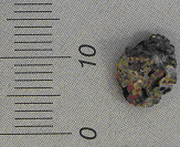
TRANSISTOR
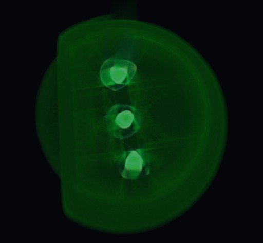
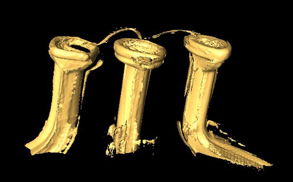
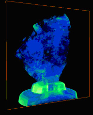
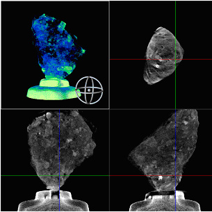
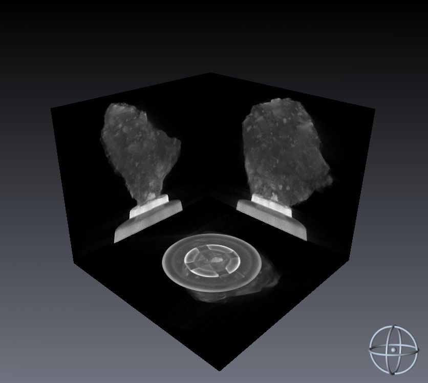
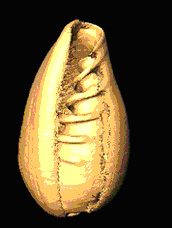
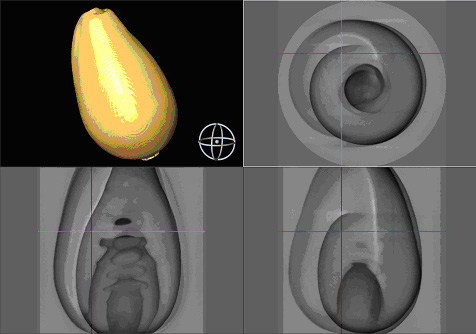
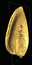
In the x-ray computed microtomography (µCT), the object is placed on a rotating base between the radiation source (X-ray tube) and the x-ray camera (detector array). The rotation is performed in steps and with each angular step the CCD x-ray sensitive camera records an x-ray image. Based on x-ray images of the object seen from different angles, a three-dimensional reconstruction of the object's interior can be made.
The following reconstructions and images were made by Jakub Bielecki on an X-ray computed microtomography facility constructed as part of his doctoral thesis at the IFJ PAN.