MICROBEAM.EU
X-ray microbeam
X-ray microbeam

at the Institute of Nuclear Physics in Krakow

Microtomography Microdosimetry

ABOUT

In 2007, at the Institute of Nuclear Physics of the Polish Academy of Sciences in Krakow, at the Department of Applied Spectroscopy, the construction of an X-ray microbeam system began. Two facilities were constructed: computed microtomography and microdosimetry. Two doctoral dissertations were carried out as part of the project. On this page you can find our publications, posters and presentations presented at conferences. Presentations of other speakers from COST group meetings Krakow and Paris are also available here.

Microtomography

X-ray computed microtomography (µCT) is a non-destructive method widely used in various disciplines. It illustrates the internal structure of the tested objects, determined by the distribution of density and atomic composition.


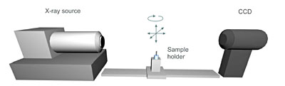
The X-ray computed microtomography method is easy to use, while ensuring high spatial resolution (about 10 µm). Visit µCT image gallery

Microdosimetry

To irradiate microscopic objects, the radiation from the source is refocused on the sample with a focusing system.


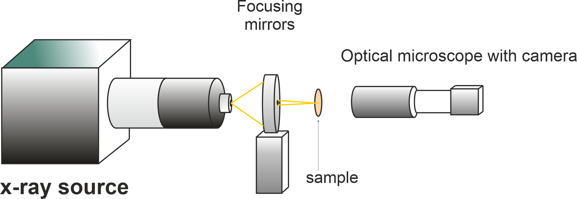
The beam diameter of the focused beam is about 20 µm, and the minimum repeatable step of the sample positioners is less than 1 µm. The beam shutter sets the precise exposure time from 120 ms. Learn more..

POSTERS

Construction and implementation of x-ray microbeam

uCT system

Investigations of elemental content, microporosity, and specific surface area 
of porous rocks using ion and X-ray microprobes

Rock porosity

x-ray microbeam resolution optimization

Resolution

x-ray targeted irradiation

Irradiation

cells irradiation complementary lines x-ray and proton microbeam

Two microbeams

x-ray microbeam single cells irradiation

Preliminary results

PRESENTATIONS

x-ray microprobe computed microtomography

Microtomography

x-ray microprobe cells irradiation

Irradiation system

PUBLICATIONS

X-ray microbeam stand-alone facility for cultured cells irradiation

S. Bożek, J. Bielecki, A. Wiecheć, J. Lekki, Z. Stachura, K. Pogoda, E. Lipiec, K. Tkocz, W. M.Kwiatek

Nuclear Instruments and Methods in Physics Research Section B: Beam Interactions with Materials and Atoms

Application of Focused X-ray Beams in Radiation Biology

J. Lekki, J. Bielecki, S. Bożek and Z. Stachura

Short Wavelength Laboratory Sources: Principles and Practices

Computed microtomography and numerical study of porous rock samples

J. Bielecki, J. Jarzyna, S. Bożek, J. Lekki, Z. Stachura, W.M. Kwiatek

Radiation Physics and Chemistry

Phase contrast imaging of lightweight objects using microfocus X-ray source and high resolution CCD camera

Z. Zaprazny, D. Korytar, V. Ac, P. Konopka, J. Bielecki,

Short Wavelength Laboratory Sources: Principles and Practices

Preliminary Investigations of Elemental Content, Microporosity, and Specific Surface Area of Porous Rocks Using PIXE and X-ray Microtomography Techniques

J. Bielecki, S. Bożek, E. Dutkiewicz, R. Hajduk, J. Jarzyna, J. Lekki, T. Pieprzyca, Z. Stachura, Z. Szklarz, W.M. Kwiatek

ACTA PHYSICA POLONICA A Vol. 121 (2012)

X-ray microbeam stand-alone facility for cultured cells irradiation

S. Bożek, J. Bielecki, A. Wiecheć, J. Lekki, Z. Stachura, K. Pogoda, E. Lipiec, K. Tkocz, W. M.Kwiatek

Nuclear Instruments and Methods in Physics Research Section B: Beam Interactions with Materials and Atoms

Experience with imaging by using of microfocus x-ray source

Z. Zaprazny, D. Korytar, F. Dubecky, V. Ac, Z. Stachura, J. Lekki, J. Bielecki, J. Mudron

Journal of ELECTRICAL ENGINEERING, VOL. 61, NO. 5, 2010, 287–290

Applications of the Cracow X-Ray Microprobe in Tomography

J. Bielecki, S. Bożek, J. Lekki, Z. Stachura, W.M. Kwiatek

ACTA PHYSICA POLONICA A, Vol. 115, 2009

X-ray microprobe - A new facility for cell irradiations in Krakow

S. Bożek, J. Bielecki, J. Baszak, , H. Doruch, R. Hajduk, J. Lekki, Z. Stachura, W.M. Kwiatek

Nuclear Instruments and Methods in Physics Research Section B, Vol. 267, 2009